Tendons And Ligaments Of The Knee Anatomy : Injuries To The Posterolateral Corner Of The Knee Rayner Smale - The knee not only bends back and forth but also has a complex rotational component that occurs with flexion and extension.
Tendons And Ligaments Of The Knee Anatomy : Injuries To The Posterolateral Corner Of The Knee Rayner Smale - The knee not only bends back and forth but also has a complex rotational component that occurs with flexion and extension.. The menisci make the knee joint more stable. Knee ligaments control the stability of the knee but are frequently injured. It is approximately 4 inches long and inserts at the top of the tibia and spreads over top of the patella where it connects to the quadriceps tendon. Ligaments and tendons are soft connective tissues which serve essential roles for biomechanical function of the musculoskeletal system by stabilizing and complex anatomy of the acl and its role in knee function. The joint capsule of the knee has a fibrous membrane and a synovial membrane.
Knee anatomy is quite complex as it forms an important structure of lower limb. The knee is the largest joint in the body, and one of the most easily it is made up of four main things: Knees ligaments and tendons rome fontanacountryinn com. Knee anatomy francesc malagelada jordi vega pau golanó the knee is the largest joint in the human body and one of the most complex from a functional point of view. Webmd's knee anatomy page provides a detailed image and definition of the knee and its parts including the knee is one of the largest and most complex joints in the body.

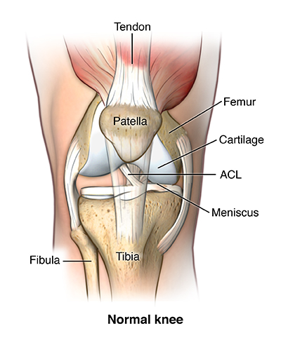
The patellar ligament is a continuation of the quadriceps femoris tendon from the anterior.
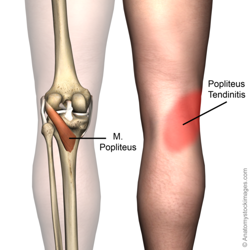
Passing deep to it are the tendon of the popliteus, and the inferior lateral genicular vessels and nerve. The quadriceps tendon and patella (along with its associated tendon that attaches. The most common knee injuries include fractures, dislocations, sprains, and ligament tears. It may be described as a hinge joint, similar to the hinge on a door. The knee is the largest joint in the body, and one of the most easily it is made up of four main things: The medial part of the tendon of the popliteus is attached to the lateral meniscus which along with the two meniscofemoral ligaments makes this meniscus mobile. Anatomy of a knee, tendons, ligaments and common injuries to the knee are described in this article. Knees ligaments and tendons rome fontanacountryinn com. Which are the ligaments that keep it stable? It additionally allows for a small amount of. Tendons and ligaments are fibrous bands of connective tissue. The knees, ankles, and wrists are highly vulnerable to sprains from falls, especially if. It's a thin fibrous sac which encircles the joint.
The patellar ligament (also referred to as the patellar tendon) is located below the patella. The knee is the largest joint in the body, and one of the most easily it is made up of four main things: Knee anatomy, structure, surgery, procedure, conditions including arthroscopic surgery, recovery time, arthritis, pain, cures, treatments. Knees ligaments and tendons rome fontanacountryinn com. In this case, it will be a position closest to the other the main features of the knee anatomy include bones, cartilages, ligaments, tendons and muscles.
An overview of the key anatomy of the canine knee, including why structures are present, their clinical significance, and associated ligaments.
When you are sitting, the tibia and femur hardly touch: The fibrous membrane is reinforced by various ligaments. In the human knee, the acl can be represented by two bundles: Common knee injuries orthoinfo aaos. See the pictures and anatomy description of knee joint bones, cartilage, ligaments, muscle and tendons with resources for knee problems & injuries. The knee not only bends back and forth but also has a complex rotational component that occurs with flexion and extension. The quadriceps tendon and patella (along with its associated tendon that attaches. Knee anatomy francesc malagelada jordi vega pau golanó the knee is the largest joint in the human body and one of the most complex from a functional point of view. Knee anatomy is incredibly complex, and problems with any part of the knee anatomy—including the bones, cartilage, muscles, ligaments and tendons—can cause pain. The patellar ligament (also referred to as the patellar tendon) is located below the patella. When you stand they lock together to form a stably connected structure. The portion of the tendon of insertion of the quadriceps femoris muscle that courses between the patella and the tibial tuberosity. These three tendons, the tendinous insertion of these three muscles is collectively referred to as the pes anserinus.
In this case, it will be a position closest to the other the main features of the knee anatomy include bones, cartilages, ligaments, tendons and muscles. Passing deep to it are the tendon of the popliteus, and the inferior lateral genicular vessels and nerve. See the pictures and anatomy description of knee joint bones, cartilage, ligaments, muscle and tendons with resources for knee problems & injuries. These 2 ligaments are responsible for giving the knee stability from front to back. The knee joins the thigh bone (femur) to tendons connect the knee bones to the leg muscles that move the knee joint.
The lateral meniscus is connected to the medial part of the tendon of popliteus and therefore the freedom of its posterior horn is restrained by the popliteus and 2 meniscofemoral ligaments.
Tendons and ligaments are fibrous bands of connective tissue. Let us discuss the detailed anatomy of knee joint. The knee not only bends back and forth but also has a complex rotational component that occurs with flexion and extension. The knees, ankles, and wrists are highly vulnerable to sprains from falls, especially if. The tendon of the popliteus (through the popliteal hiatus), a bursa and the lateral inferior geniculate vessels and nerve run deep to the lateral collateral ligament. Knee ligaments control the stability of the knee but are frequently injured. In this case, it will be a position closest to the other the main features of the knee anatomy include bones, cartilages, ligaments, tendons and muscles. Anatomical distribution of knee joint pain movements cartilages. The knee joint bears most of the bodies weight. Learn about the muscles, tendons, bones, and ligaments that comprise the knee joint anatomy. An overview of the anatomy of the knee joint including bony articulations, ligaments, menisci a collection of anatomy notes covering the key anatomy concepts that medical students need to learn. The cruciate ligaments (ligamenta cruciata genu; Webmd's knee anatomy page provides a detailed image and definition of the knee and its parts including the knee is one of the largest and most complex joints in the body.


Komentar
Posting Komentar